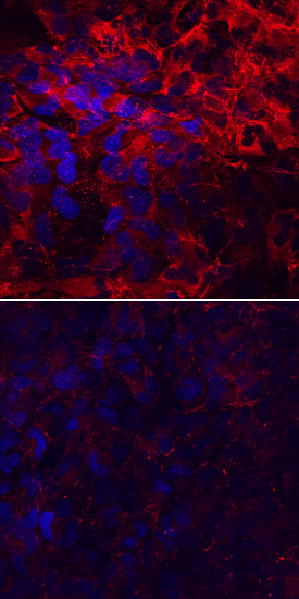
Laminin alpha 4 was detected in immersion fixed T98G human glioblastoma cell line, induced (upper panel) or not (lower panel) with StemXVivo EMT Inducing Media Supplement (Catalog # CCM017), using Sheep Anti-Human Laminin alpha 4 Antigen Affinity-purified Polyclonal Antibody (Catalog # AF7340) at 10 µg/mL for 3 hours at room temperature. Cells were stained using the NorthernLights™ 557-conjugated Anti-Sheep IgG Secondary Antibody (red; Catalog # NL010) and counterstained with DAPI (blue). Specific staining was localized to cytoplasm and cell surfaces. View our protocol for Fluorescent ICC Staining of Cells on Coverslips.

Western blot shows lysates of human placenta tissue. PVDF membrane was probed with 2 µg/mL of Sheep Anti-Human Laminin alpha 4 Antigen Affinity-purified Polyclonal Antibody (Catalog # AF7340) followed by HRP-conjugated Anti-Sheep IgG Secondary Antibody (Catalog # HAF016). A specific band was detected for Laminin alpha 4 at approximately 200‑220 kDa (as indicated). This experiment was conducted under reducing conditions and using Immunoblot Buffer Group 1.

Recombinant Human Laminin alpha 4 (Catalog # 7340-A4) supports adhesion in the HT1080 human fibrosarcoma cell line in a dose-dependent manner (orange line), as measured by Calcein AM. Adhesion elicited by Recombinant Human Laminin alpha 4 (5 µg/mL) is neutralized (green line) by increasing concentrations of Sheep Anti-Human Laminin alpha 4 Antigen Affinity-purified Polyclonal Antibody (Catalog # AF7340). The ND50 is typically1.5-7.5 µg/mL.