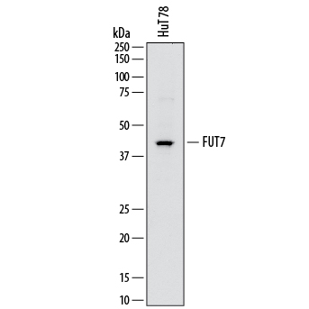
Western blot shows lysates of HuT 78 human cutaneous T cell lymphoma cell line. PVDF membrane was probed with 2 µg/mL of Mouse Anti-Human Fucosyltransferase 7/FUT7 Monoclonal Antibody (Catalog # MAB64091) followed by HRP-conjugated Anti-Mouse IgG Secondary Antibody (Catalog # HAF018). A specific band was detected for Fucosyltransferase 7/FUT7 at approximately 45 kDa (as indicated). This experiment was conducted under reducing conditions and using Immunoblot Buffer Group 1.

Fucosyltransferase 7/FUT7 was detected in immersion fixed HL‑60 human acute promyelocytic leukemia cell line using Mouse Anti-Human Fucosyltransferase 7/FUT7 Monoclonal Antibody (Catalog # MAB64091) at 10 µg/mL for 3 hours at room temperature. Cells were stained using the NorthernLights™ 557-conjugated Anti-Mouse IgG Secondary Antibody (red; Catalog # NL007) and counterstained with DAPI (blue). Specific staining was localized to cytoplasm. View our protocol for Fluorescent ICC Staining of Non-adherent Cells.