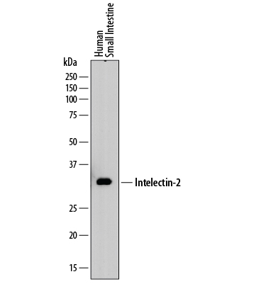
Western blot shows lysates of human small intestine tissue. PVDF membrane was probed with 2 µg/mL of Mouse Anti-Human Intelectin‑2 Monoclonal Antibody (Catalog # MAB8004) followed by HRP-conjugated Anti-Mouse IgG Secondary Antibody (Catalog # HAF018). A specific band was detected for Intelectin‑2 at approximately 35 kDa (as indicated). This experiment was conducted under reducing conditions and using Immunoblot Buffer Group 1.

Intelectin‑2 was detected in formalin fixed paraffin-embedded sections of human small intestine using Mouse Anti-Human Intelectin‑2 Monoclonal Antibody (Catalog # MAB8004) at 5 µg/mL overnight at 4 °C. Tissue was stained using the Anti-Mouse HRP-DAB Cell & Tissue Staining Kit (brown; Catalog # CTS002) and counterstained with hematoxylin (blue). Specific staining was localized to Paneth cells in intestinal glands. View our protocol for Chromogenic IHC Staining of Paraffin-embedded Tissue Sections.