
Western blot shows lysates of human brain (hippocampus) tissue, mouse brain tissue, and rat brain (cerebellum) tissue. PVDF membrane was probed with 0.5 µg/mL of Goat Anti-Human/Mouse/Rat Complexin-1/2 Antigen Affinity-purified Polyclonal Antibody (Catalog # AF7787) followed by HRP-conjugated Anti-Goat IgG Secondary Antibody (Catalog # HAF017). Specific bands were detected for Complexin-1/2 at approximately 17-19 kDa (as indicated). This experiment was conducted under reducing conditions and using Immunoblot Buffer Group 1.
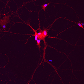
Complexin-1/2 was detected in immersion fixed rat hippocampal neurons (14 days in vitro) using Goat Anti-Human/Mouse/Rat Complexin-1/2 Antigen Affinity-purified Polyclonal Antibody (Catalog # AF7787) at 10 µg/mL for 3 hours at room temperature. Cells were stained using the NorthernLights™ 557-conjugated Anti-Goat IgG Secondary Antibody (red; Catalog # NL001) and counterstained with DAPI (blue). Specific staining was localized to cytoplasm. View our protocol for Fluorescent ICC Staining of Cells on Coverslips.