
Western blot shows lysates of mouse, human, and rat brain tissue. PVDF membrane was probed with 1 µg/mL of Goat Anti-Human/Mouse/Rat Complexin‑2 Antigen Affinity-purified Polyclonal Antibody (Catalog # AF5085) followed by HRP-conjugated Anti-Goat IgG Secondary Antibody (Catalog # HAF109). A specific band was detected for Complexin‑2 at approximately 19 kDa (as indicated). This experiment was conducted under reducing conditions and using Immunoblot Buffer Group 2.
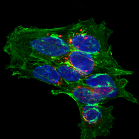
SH‑SY5Y human neuroblastoma cells were cultured overnight in the presence of 1 mM Retinoic Acid (Catalog # 0695/50) prior to immersion fixation. Complexin-2 was detected using a Goat Anti-Human/Mouse/Rat Complexin-2 Antigen Affinity-purified Polyclonal Antibody (Catalog # AF5085). The cells were stained with the NorthernLights 557-conjugated Donkey Anti-Goat IgG Affinity-purified Secondary Antibody (red; Catalog # NL001). Actin filaments were stained with FITC-conjugated Phalloidin (green) and the nuclei were counterstained with DAPI (blue). Complexin-2 immunoreactivity was localized to synaptic vesicles. View our protocol for Fluorescent ICC Staining of Cells on Coverslips.