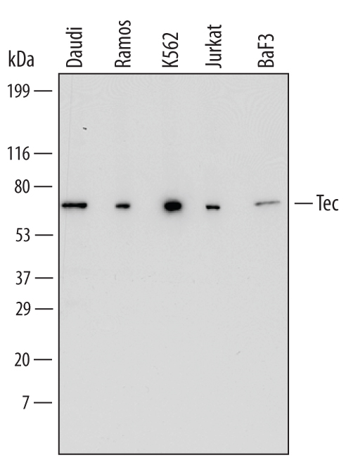
Western blot shows lysates of Daudi human Burkitt's lymphoma cell line, Ramos human Burkitt's lymphoma cell line, K562 human chronic myelogenous leukemia cell line, Jurkat human acute T cell leukemia cell line, and BaF3 mouse pro-B cell line. PVDF Membrane was probed with 1 µg/mL of Human/Mouse Tec Monoclonal Antibody (Catalog # MAB6519) followed by HRP-conjugated Anti-Mouse IgG Secondary Antibody (Catalog # HAF007). A specific band was detected for Tec at approximately 66 kDa (as indicated). This experiment was conducted under reducing conditions and using Immunoblot Buffer Group 1.
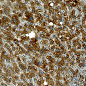
Tec was detected in immersion fixed paraffin-embedded sections of human liver using Human/Mouse Tec Monoclonal Antibody (Catalog # MAB6519) at 15 µg/mL overnight at 4 °C. Before incubation with the primary antibody, tissue was subjected to heat-induced epitope retrieval using Antigen Retrieval Reagent-Basic (Catalog # CTS013). Tissue was stained using the Anti-Mouse HRP-DAB Cell & Tissue Staining Kit (brown; Catalog # CTS002) and counterstained with hematoxylin (blue). Specific staining was localized to cytoplasm in hepatocytes. View our protocol for Chromogenic IHC Staining of Paraffin-embedded Tissue Sections.