
Western blot shows lysates of NCI‑H460 human large cell lung carcinoma cell line. PVDF Membrane was probed with 2 µg/mL of Human Collagen XXIII alpha 1 Monoclonal Antibody (Catalog # MAB4165) followed by HRP-conjugated Anti-Mouse IgG Secondary Antibody (Catalog # HAF007). Specific bands were detected for Collagen XXIII alpha 1 at approximately 75 and 60 kDa (as indicated). This experiment was conducted under reducing conditions and using Immunoblot Buffer Group 1.
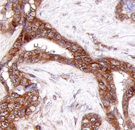
Collagen XXIII alpha 1 was detected in immersion fixed paraffin-embedded sections of human adenocarcinoma using Human Collagen XXIII alpha 1 Monoclonal Antibody (Catalog # MAB4165) at 25 µg/mL overnight at 4 °C. Tissue was stained using the Anti-Mouse HRP-DAB Cell & Tissue Staining Kit (brown; Catalog # CTS002) and counterstained with hematoxylin (blue). Specific staining was localized to cytoplasm. View our protocol for Chromogenic IHC Staining of Paraffin-embedded Tissue Sections.