
Western blot shows recombinant human Syntaxin 12, 16, 1A, 1B2, 5, 6, 7, 8, and 1B1 (5 ng/lane). PVDF membrane was probed with 1 µg/mL Sheep Anti-Human/Mouse/Rat Syntaxin 8 Antigen Affinity-purified Polyclonal Antibody (Catalog # AF5448) followed by HRP-conjugated Anti-Sheep IgG Secondary Antibody (Catalog # HAF016). A specific band for Syntaxin 8 was detected as indicated. This experiment was conducted under reducing conditions and using Immunoblot Buffer Group 1.

Western blot shows lysates of HeLa human cervical epithelial carcinoma cell line, Jurkat human acute T cell leukemia cell line, L1.2 mouse pro-B cell line, and Rat-2 rat embryonic fibroblast cell line. PVDF membrane was probed with 1 µg/mL Sheep Anti-Human/Mouse/Rat Syntaxin 8 Antigen Affinity-purified Polyclonal Antibody (Catalog # AF5448) followed by HRP-conjugated Anti-Sheep IgG Secondary Antibody (Catalog # HAF016). A specific band for Syntaxin 8 was detected at approximately 28 kDa (as indicated). This experiment was conducted under reducing conditions and using Immunoblot Buffer Group 1.
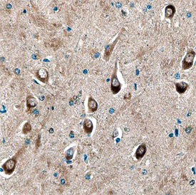
Syntaxin 8 was detected in immersion fixed paraffin-embedded sections of human brain using Sheep Anti-Human/Mouse/Rat Syntaxin 8 Antigen Affinity-purified Polyclonal Antibody (Catalog # AF5448) at 10 µg/mL overnight at 4 °C. Before incubation with the primary antibody, tissue was subjected to heat-induced epitope retrieval using Antigen Retrieval Reagent-Basic (Catalog # CTS013). Tissue was stained using the Anti-Sheep HRP-DAB Cell & Tissue Staining Kit (brown; Catalog # CTS019) and counterstained with hematoxylin (blue). Specific staining was localized to neuronal cell bodies and processes. View our protocol for Chromogenic IHC Staining of Paraffin-embedded Tissue Sections.