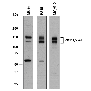
Western blot shows lysates of MO7e human megakaryocytic leukemic cell line, P815 mouse mastocytoma cell line, and MC/9‑2 mouse mast cell line. PVDF membrane was probed with 0.1 µg/mL of Goat Anti-Human/Mouse CD117/c-kit Antigen Affinity-purified Polyclonal Antibody (Catalog # AF1356) followed by HRP-conjugated Anti-Goat IgG Secondary Antibody (Catalog # HAF017). Specific bands were detected for CD117/c-kit at approximately 135, 150 kDa (as indicated). This experiment was conducted under reducing conditions and using Immunoblot Buffer Group 1.
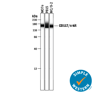
Simple Western lane view shows lysates of MO7e human megakaryocytic leukemic cell line, P815 mouse mastocytoma cell line and MC/9-2 mouse mast cell line, loaded at 0.2 mg/mL. A specific band was detected for CD117/c-kit at approximately 150-165 kDa (as indicated) using 5 µg/mL of Goat Anti-Human/Mouse CD117/c-kit Antigen Affinity-purified Polyclonal Antibody (Catalog # AF1356) followed by 1:50 dilution of HRP-conjugated Anti-Goat IgG Secondary Antibody (Catalog # HAF109). This experiment was conducted under reducing conditions and using the 12-230 kDa separation system.
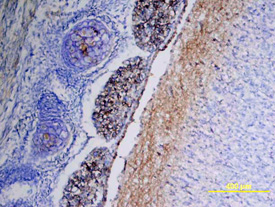
CD117/c-kit was detected in immersion fixed frozen sections of mouse embryo using Goat Anti-Human/Mouse CD117/c-kit Antigen Affinity-purified Polyclonal Antibody (Catalog # AF1356) at 15 µg/mL overnight at 4 °C. Tissue was stained using the Anti-Goat HRP-DAB Cell & Tissue Staining Kit (brown; Catalog # CTS008) and counterstained with hematoxylin (blue). View our protocol for Chromogenic IHC Staining of Frozen Tissue Sections.