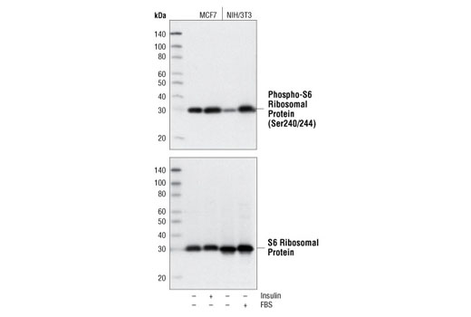
Western blot analysis of extracts from MCF7 and NIH/3T3 cells, treated with 100 nM insulin (10 min) or 20% FBS (30 min) as indicated, using Phospho-S6 Ribosomal Protein (Ser240/244) (D68F8) XP ® Rabbit mAb (upper) or S6 Ribosomal Protein (5G10) Rabbit mAb #2217 (lower).
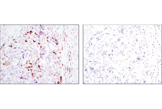
Immunohistochemical analysis of paraffin-embedded human colon carcinoma using Phospho-S6 Ribosomal Protein (Ser240/244) (D68F8) XP ® Rabbit mAb in the presence of control peptide (left) or antigen-specific peptide (right).
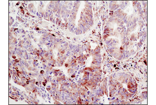
Immunohistochemical analysis of paraffin-embedded human lung carcinoma using Phospho-S6 Ribosomal Protein (Ser240/244) (D68F8) XP ® Rabbit mAb.

Immunohistochemical analysis of paraffin-embedded mouse colon using Phospho-S6 Ribosomal Protein (Ser240/244) (D68F8) XP ® Rabbit mAb.

Immunohistochemical analysis on SignalSlide ® Phospho-Akt (Ser473) IHC Controls #8101 (paraffin-embedded LNCaP cell pellets -/+ LY294002) using Phospho-S6 Ribosomal Protein (Ser240/244) (D68F8) XP ® Rabbit mAb.

Immunohistochemical analysis of paraffin-embedded LNCaP cell pellets, control (left) or rapamycin-treated (right), using Phospho-S6 Ribosomal Protein (Ser240/244) (D68F8) XP ® Rabbit mAb.
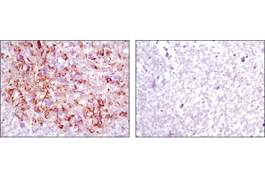
Immunohistochemical analysis of paraffin-embedded Rh30 xenograft, control (left) or rapamycin-treated (right), using Phospho-S6 Ribosomal Protein (Ser240/244) (D68F8) XP ® Rabbit mAb.
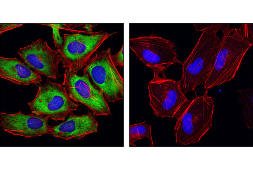
Confocal immunofluorescent analysis of HeLa cells, insulin-treated (left) and LY294002-treated (#9901, right), using Phospho-S6 Ribosomal Protein (Ser240/244) (D68F8) XP ® Rabbit mAb (green). Actin filaments were labeled with DY-554 phalloidin (red). Blue pseudocolor = DRAQ5 ® #4084 (fluorescent DNA dye).
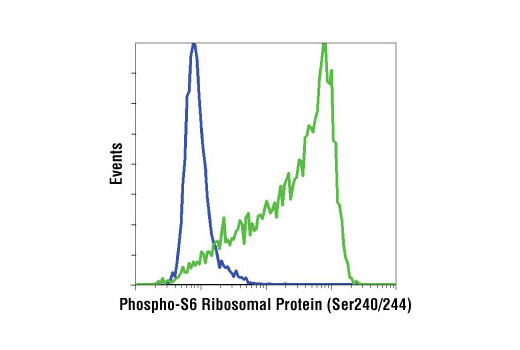
Flow cytometric analysis of Jurkat cells, untreated (green) or treated with LY294002, wortmannin and U0126 (blue), using Phospho-S6 Ribosomal Protein (Ser240/244) (D68F8) XP ® Rabbit mAb.