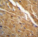
TBC1D13 Antibody immunohistochemistry of formalin-fixed and paraffin-embedded mouse heart tissue followed by peroxidase-conjugated secondary antibody and DAB staining.

Confocal immunofluorescent of TBC1D13 Antibody with NCI-H460 cell followed by Alexa Fluor 488-conjugated goat anti-rabbit lgG (green). Actin filaments have been labeled with Alexa Fluor 555 phalloidin (red). DAPI was used to stain the cell nuclear (blue).

TBC1D13 Antibody western blot of CEM cell line lysates (35 ug/lane). The TBC1D13 antibody detected the TBC1D13 protein (arrow).
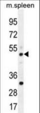
TBC1D13 Antibody western blot of mouse spleen tissue lysates (35 ug/lane). The TBC1D13 antibody detected the TBC1D13 protein (arrow).