RARγ1 (D3A4) XP ® Rabbit mAb
| Name | RARγ1 (D3A4) XP ® Rabbit mAb |
|---|---|
| Supplier | Cell Signaling Technology |
| Catalog | 8965 |
| Prices | $109.00, $277.00 |
| Sizes | 20 µl (2 western blots), 100 µl (10 western blots) |
| Host | Rabbit |
| Clonality | Monoclonal |
| Isotype | IgG |
| Clone | D3A4 |
| Applications | WB IP IHC-P ICC/IF |
| Species Reactivities | Human, Mouse, Rat, Hamster, Bovine, Dog |
| Antigen | Monoclonal antibody is produced by immunizing animals with a synthetic peptide corresponding to residues near the amino terminus of human RARγ1 protein. |
| Description | Rabbit Monoclonal |
| Gene | RARG |
| Supplier Page | Shop |
Product images
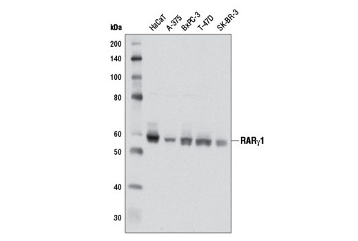 Western blot analysis of extracts from various cell lines using RARγ1 (D3A4) XP ® Rabbit mAb.
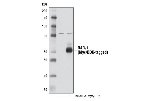 Western blot analysis of extracts from 293T cells, either mock transfected (-) or transfected with a Myc/DDK-tagged cDNA expression construct encoding full-length human RARγ1 (hRARγ1-Myc/DDK, +), using RARγ1 (D3A4) XP ® Rabbit mAb.
 Immunohistochemical analysis of paraffin-embedded cell pellets, HaCaT (positive, left) and Hep3B (negative, right), using RARγ1 (D3A4) XP ® Rabbit mAb.
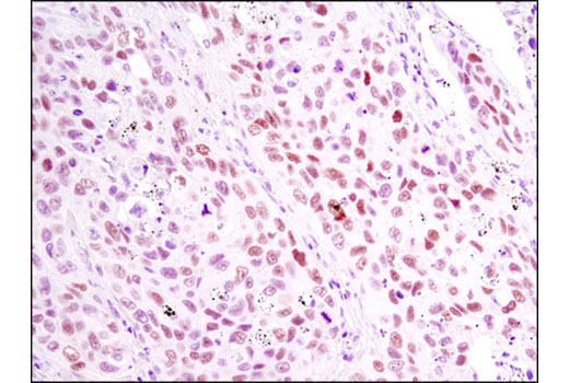 Immunohistochemical analysis of paraffin-embedded human lung carcinoma using RARγ1 (D3A4) XP ® Rabbit mAb.
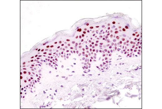 Immunohistochemical analysis of paraffin-embedded human skin using RARγ1 (D3A4) XP ® Rabbit mAb.
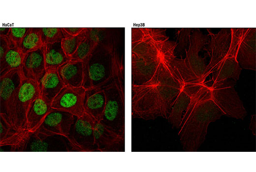 Confocal immunofluorescent analysis of HaCaT cells (positive, left) and Hep3B cells (negative, right) using RARγ1 (D3A4) XP ® Rabbit mAb (green). Actin filaments were labeled with DY-554 phalloidin (red).