Anti-SREBF1 / SREBP-1 Antibody (aa349-378)
| Name | Anti-SREBF1 / SREBP-1 Antibody (aa349-378) |
|---|---|
| Supplier | LifeSpan Bioscience |
| Catalog | LS-C161494 |
| Prices | $295.00 |
| Sizes | 400 µl |
| Host | Rabbit |
| Clonality | Polyclonal |
| Applications | IHC-P WB |
| Species Reactivities | Human, Mouse, Rat, Bovine, Goat, Pig, Sheep |
| Purity/Format | Protein A purified |
| Blocking Peptide | SREB / GPR85 Antibody Blocking Peptide |
| Description | Rabbit Polyclonal |
| Gene | SREBF1 |
| Conjugate | Unconjugated |
| Supplier Page | Shop |
Product images
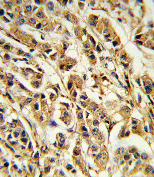 Formalin-fixed and paraffin-embedded human breast carcinoma reacted with SREBF1 Antibody , which was peroxidase-conjugated to the secondary antibody, followed by DAB staining. This data demonstrates the use of this antibody for immunohistochemistry; clinical relevance has not been evaluated.
 Western blot of SREBF1 Antibody in MDA-MB435 cell line lysates (35 ug/lane). SREBF1 (arrow) was detected using the purified antibody.
 SREBF1 Antibody flow cytometry of Ramos cells (bottom histogram) compared to a negative control cell (top histogram). FITC-conjugated goat-anti-rabbit secondary antibodies were used for the analysis.