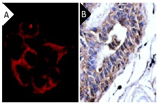
NCK2 (8.8): sc-20020. Immunofluorescence staining of methanol-fixed H4 cells (A) and immunoperoxidase staining of formalin-fixed, paraffin-embedded human breast tumor (B) showing cytoplasmic staining.

NCK2 (8.8): sc-20020. Western blot analysis of NCK2 expression in U-937 whole cell lysate.
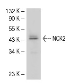
NCK2 (8.8): sc-20020. Western blot analysis of NCK2 expression in U-937 whole cell lysate.
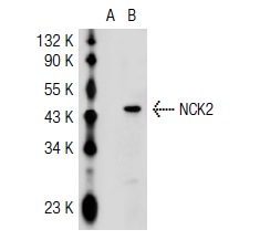
NCK2 (8.8): sc-20020. Western blot analysis of NCK2 expression in non-transfected: sc-117752 (A) and mouse NCK2 transfected: sc-121954 (B) 293T whole cell lysates.
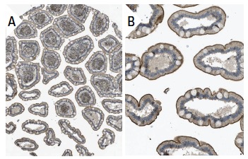
NCK2 (8.8): sc-20020. Immunoperoxidase staining of formalin fixed, paraffin-embedded human small intestine tissue showing membrane staining of microvilli in glandular cells at low (A) and high (B) magnification. Kindly provided by The Swedish Human Protein Atlas (HPA) program.
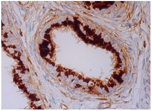
NCK2 (8.8): sc-20020. Immunoperoxidase staining of formalin fixed, paraffin-embedded human epididymis tissue showing cytoplasmic staining of glandular cells.