PRNP (D3Q5C) Rabbit mAb
| Name | PRNP (D3Q5C) Rabbit mAb |
|---|---|
| Supplier | Cell Signaling Technology |
| Catalog | 14025 |
| Prices | $246.00 |
| Sizes | 100 µl (10 western blots) |
| Host | Rabbit |
| Clonality | Monoclonal |
| Isotype | IgG |
| Clone | D3Q5C |
| Applications | WB IP ICC/IF |
| Species Reactivities | Human, Mouse, Rat |
| Antigen | Monoclonal antibody is produced by immunizing animals with a synthetic peptide corresponding to residues surrounding Ser222 of human PRNP protein. |
| Description | Rabbit Monoclonal |
| Gene | PRNP |
| Supplier Page | Shop |
Product images
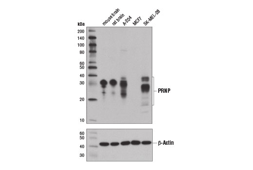 Western blot analysis of extracts from various tissues and cell lines using PRNP (D3Q5C) Rabbit mAb (upper) and β-Actin (D6A8) Rabbit mAb #8457 (lower).
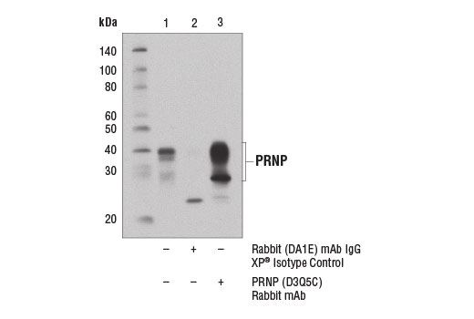 Immunoprecipitation of PRNP from SK-MEL-28 cell extracts using Rabbit (DA1E) mAb IgG XP ® Isotype Control #3900 (lane 2) or PRNP (D3Q5C) Rabbit mAb (lane 3). Lane 1 is 10% input. Western blot analysis was performed using PRNP (D3Q5C) Rabbit mAb.
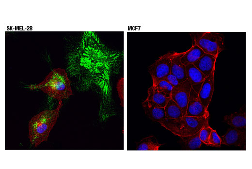 Confocal immunofluorescent analysis of SK-MEL-28 (positive, left) or MCF7 (negative, right) cells, using PRNP (D3Q5C) Rabbit mAb (green). Actin filaments were labeled with DyLight™ 554 Phalloidin #13054 (red). Blue pseudocolor = DRAQ5 ® #4084 (fluorescent DNA dye).