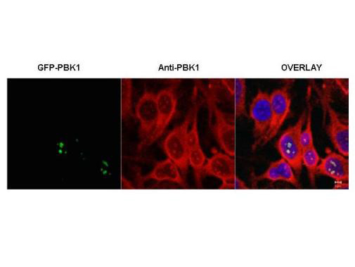
Anti-PBK1 Antibody - Immunofluorescence Microscopy. Immunofluorescence microscopy of HeLa cells transfected with GFP-PBK1. In the overlay, specific antibody staining is shown to co-localize with recombinant protein. Cells were fixed with methanol prior to staining. Personal communication, J. McNally and D. Stavreva, NCI, Bethesda, MD.
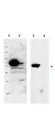
Anti-PBK1 Antibody - Western Blot. Western blot of affinity purified anti-PBK1 antibody shows detection of over-expressed PBK1 in lysates from HeLa cells transfected with Flag-PBK1. Lanes 1 and 3 contain lysate from Flag-PBK1 transfected HeLa cells. Lanes 2 and 4 contain lysate from cells transfected with null vector. Lanes 1 and 2 were blotted with anti-Flag antibody. Lanes 3 and 4 were probed with a 1:500 dilution of anti-PBK1. The band at 75 kD, indicated by the arrowhead, corresponds to PBK1. Personal communication, J. McNally and D. Stavreva, NCI, Bethesda, MD.