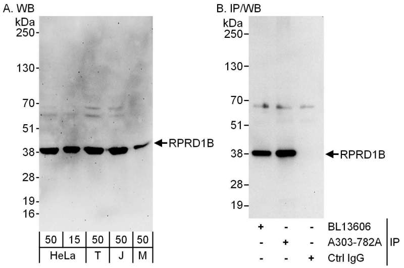
Detection of Human and Mouse RPRD1B by Western Blot (h and m) and Immunoprecipitation (h). Samples: Whole cell lysate from HeLa (15 and 50 ug for WB; 1 mg for IP, 20% of IP loaded), 293T (T; 50 ug), Jurkat (J; 50 ug) and mouse NIH3T3 (M; 50 ug) cells. Antibodies: Affinity purified rabbit anti-RPRD1B antibody used for WB at 0.1 ug/ml (A) and 1 ug/ml (B) and used for IP at 6 ug/mg lysate. RPRD1B was also immunoprecipitated by rabbit anti-RPRD1B antibody BL13606, which recognizes an upstream epitope. Detection: Chemiluminescence with exposure times of 3 minutes (A) and 10 seconds (B).