Anti-RNF208 Antibody (aa86-115)
| Name | Anti-RNF208 Antibody (aa86-115) |
|---|---|
| Supplier | LifeSpan Bioscience |
| Catalog | LS-C162656 |
| Prices | $295.00 |
| Sizes | 400 µl |
| Host | Rabbit |
| Clonality | Polyclonal |
| Applications | IHC-P WB FC |
| Species Reactivities | Human, Mouse |
| Purity/Format | Protein A purified |
| Blocking Peptide | RNF19A / DORFIN Antibody Blocking Peptide |
| Description | Rabbit Polyclonal |
| Gene | RNF208 |
| Conjugate | Unconjugated |
| Supplier Page | Shop |
Product images
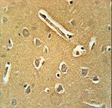 RNF208 Antibody immunohistochemistry of formalin-fixed and paraffin-embedded human brain tissue followed by peroxidase-conjugated secondary antibody and DAB staining.
 Western blot of lysate from A431 cell line, using RNF208 Antibody. Antibody was diluted at 1:1000 at each lane. A goat anti-rabbit IgG H&L (HRP) at 1:5000 dilution was used as the secondary antibody. Lysate at 35ug per lane.
 RNF208 Antibody flow cytometry of HepG2 cells (bottom histogram) compared to a negative control cell (top histogram). FITC-conjugated goat-anti-rabbit secondary antibodies were used for the analysis.