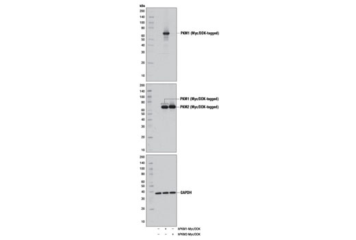
Western blot analysis of extracts from 293 cells, mock transfected (-) or transfected with a construct expressing Myc/DDK-tagged full-length human PKM1 (hPKM1-Myc/DDK; +) or Myc/DDK-tagged full-length human PKM2 (hPKM2-Myc/DDK; +), using PKM1 (D30G6) XP ® Rabbit mAb (upper), DYKDDDDK (9A3) Mouse mAb #8146 (middle), or GAPDH (D16H11) XP ® Rabbit mAb #5174 (lower).

Western blot analysis of extracts from various mouse and human tissues using PKM1 (D30G6) XP ® Rabbit mAb (upper) or GAPDH (D16H11) XP ® Rabbit mAb #5174 (lower).

Immunohistochemical analysis of paraffin-embedded mouse liver using PKM1 (D30G6) XP ® Rabbit mAb. Note the staining of vascular smooth muscle with no staining of hepatocytes.

Immunohistochemical analysis of paraffin-embedded human colon using PKM1 (D30G6) XP ® Rabbit mAb in the presence of control peptide (left) or antigen-specific peptide (right).

Confocal immunofluorescent analysis of mouse skeletal muscle (left) or liver (right) using PKM1 (D30G6) XP ® Rabbit mAb (green). Blue pseudocolor = DRAQ5 ® #4084 (fluorescent DNA dye).
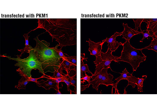
Confocal immunofluorescent analysis of COS-7 cells, transiently transfected with PKM1 (left) or PKM2 (right), using PKM1 (D30G6) XP ® Rabbit mAb (green). Actin filaments were labeled with DyLight™ 554 Phalloidin #13054 (red). Blue pseudocolor = DRAQ5 ® #4084 (fluorescent DNA dye).