Anti-REG3G Antibody (aa89-116) IHC-plusâ¢
| Name | Anti-REG3G Antibody (aa89-116) IHC-plus⢠|
|---|---|
| Supplier | LifeSpan Bioscience |
| Catalog | LS-B10435 |
| Prices | $395.00 |
| Sizes | 200 µl |
| Host | Rabbit |
| Clonality | Polyclonal |
| Applications | IHC WB FC ELISA |
| Species Reactivities | Human, Mouse |
| Purity/Format | Protein A purified |
| Blocking Peptide | REEP5 Antibody Blocking Peptide |
| Description | Rabbit Polyclonal |
| Gene | REG3G |
| Conjugate | Unconjugated |
| Supplier Page | Shop |
Product images
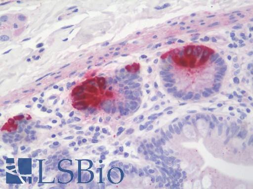 Anti-REG3G antibody IHC staining of human small intestine. Immunohistochemistry of formalin-fixed, paraffin-embedded tissue after heat-induced antigen retrieval. Antibody LS-B10435 dilution 1:100.
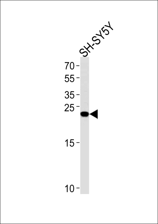 REG3G Antibody western blot of SH-SY5Y cell line lysates (35 ug/lane). The REG3G antibody detected the REG3G protein (arrow).
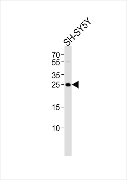 Western blot of lysate from SH-SY5Y cell line, using REG3G Antibody. Antibody was diluted at 1:1000. A goat anti-rabbit IgG H&L (HRP) at 1:5000 dilution was used as the secondary antibody. Lysate at 35ug.
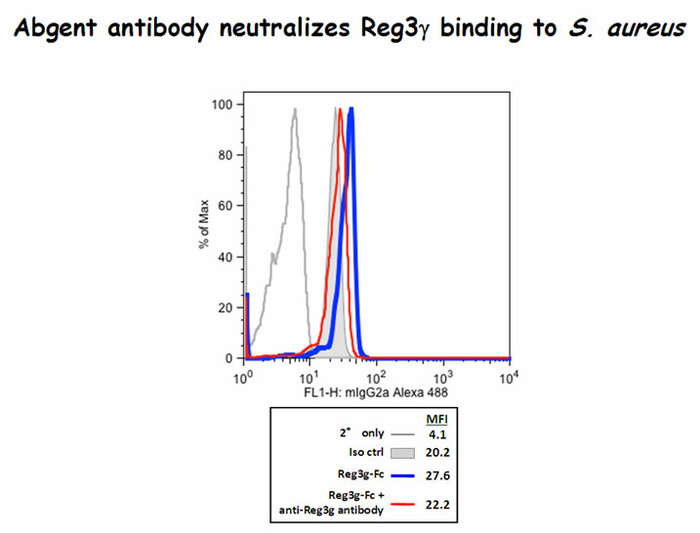 Reg3g binds to Staphylococcus aureus and the antibody did block some of this binding(Kindly offered by Dr. Choi).