PFKFB2 (D7G5R) Rabbit mAb
| Name | PFKFB2 (D7G5R) Rabbit mAb |
|---|---|
| Supplier | Cell Signaling Technology |
| Catalog | 13045 |
| Prices | $246.00 |
| Sizes | 100 µl (10 western blots) |
| Host | Rabbit |
| Clonality | Monoclonal |
| Isotype | IgG |
| Clone | D7G5R |
| Applications | WB IP ICC/IF |
| Species Reactivities | Human, Monkey |
| Antigen | Monoclonal antibody is produced by immunizing animals with a synthetic peptide corresponding to residues surrounding Pro454 within the fructose-2,6-biphosphatase region of human PFKFB2 protein. |
| Description | Rabbit Monoclonal |
| Gene | PFKFB2 |
| Supplier Page | Shop |
Product images
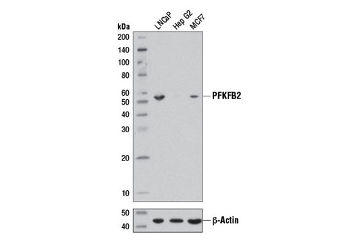 Western blot analysis of extracts from LNCaP, Hep G2, and MCF7 cells using PFKFB2 (D7G5R) Rabbit mAb (upper) and β-Actin (D6A8) Rabbit mAb #8457 (lower).
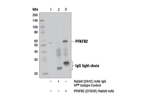 Immunoprecipitation of PFKFB2 from Hep G2 cell extracts using Rabbit (DA1E) mAb IgG XP ® Isotype Control #3900 (lane 2) or PFKFB2 (D7G5R) Rabbit mAb (lane 3). Lane 1 is 10% input. Western blot analysis was performed using PFKFB2 (D7G5R) Rabbit mAb. An anti-rabbit IgG light chain antibody was used as the secondary antibody.
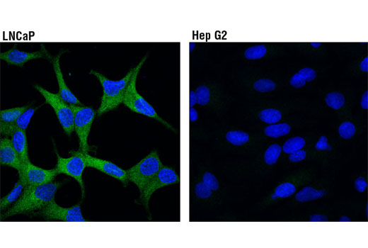 Confocal immunofluorescent analysis of LNCaP (high expression, left) and Hep G2 (low expression, right) cells using PFKFB2 (D7G5R) Rabbit mAb (green). Blue pseudocolor = DRAQ5 ® #4084 (fluorescent DNA dye).