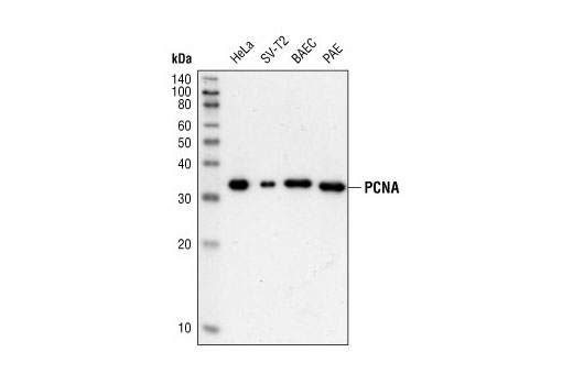
Western blot analysis of human (HeLa), murine (SV-T2), bovine (BAEC), and porcine (PAE) cell extracts using PCNA (PC10) Mouse mAb.

Western blot analysis, using PCNA (PC10) Mouse mAb (upper) and Beta-Actin Antibody #4967(lower), of HeLa cell extract (lane 1) and PCNA immunoprecipitated from the same extract using PCNA (PC10) Mouse mAb (lane 2).

Immunohistochemical analysis of paraffin-embedded human breast carcinoma, showing nuclear localization, using PCNA (PC10) Mouse mAb.

Immunohistochemical analysis of paraffin-embedded human colon carcinoma, showing nuclear localization, using PCNA (PC10) Mouse mAb.

Immunohistochemical analysis of paraffin-embedded human Non-Hodgkin's lymphoma, showing nuclear localization, using PCNA (PC10) Mouse mAb.

Confocal immunofluorescent analysis of NCI-H460 cells using PCNA (PC10) Mouse mAb (green), Phospho-Histone H3 (Ser10) (D2C8) XP Rabbit mAb (Alexa Fluor 555 Conjugate) (red), and p21 Waf1/Cip1 (12D1) Rabbit mAb (blue).

Flow cytometric analysis of Jurkat cells using PCNA (PC10) Mouse mAb and Propidium Iodide (PI)/RNase Staining Solution #4087 to measure DNA content. Anti-Mouse IgG (H+L), F(ab’) 2 Fragment (Alexa Fluor ® 488 Conjugate) #4408 was used as a secondary antibody.