PAK1 Antibody
| Name | PAK1 Antibody |
|---|---|
| Supplier | Cell Signaling Technology |
| Catalog | 2602 |
| Prices | $99.00, $246.00 |
| Sizes | 20 µl (2 western blots), 100 µl (10 western blots) |
| Host | Rabbit |
| Clonality | Polyclonal |
| Applications | WB IP IHC-P |
| Species Reactivities | Human, Mouse, Rat, Monkey, Guinea Pig |
| Antigen | Polyclonal antibodies are produced by immunizing animals with a synthetic peptide corresponding to the amino-terminus of human PAK1 |
| Description | Rabbit Polyclonal |
| Gene | PAK1 |
| Supplier Page | Shop |
Product images
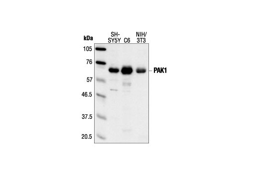 Western blot analysis of extracts from SH-SY5Y, C6 and NIH/3T3 cells, using PAK1 Antibody.
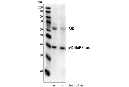 Western blot analysis of extracts from HeLa cells transfected with 100 nM control siRNA #6201 (-) or PAK1 siRNA, using PAK1 Antibody #2602 and p42 MAP Kinase (Erk2) Antibody #9108. The PAK1 Antibody confirms silencing of PAK1 expression, and p42 MAP Kinase (Erk2) Antibody is used to control for loading and specificity of PAK1 siRNA.
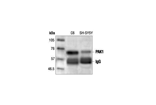 Immunoprecipitation of PAK1 from C6 and SH-SY5Y cells followed by Western blot analysis, using PAK1 Antibody.
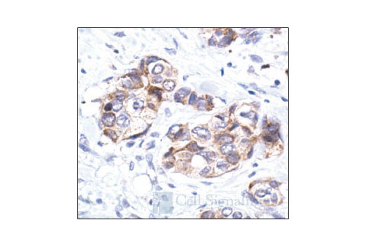 Immunohistochemical analysis of paraffin-embedded human breast carcinoma, showing cytoplasmic localization, using PAK1 Antibody.