Anti-PGD Antibody (aa236-265)
| Name | Anti-PGD Antibody (aa236-265) |
|---|---|
| Supplier | LifeSpan Bioscience |
| Catalog | LS-C163549 |
| Prices | $295.00 |
| Sizes | 400 µl |
| Host | Rabbit |
| Clonality | Polyclonal |
| Applications | IHC ICC/IF WB ELISA |
| Species Reactivities | Yeast |
| Purity/Format | Protein A purified |
| Blocking Peptide | PGAP3 / PERLD1 Antibody Blocking Peptide |
| Description | Rabbit Polyclonal |
| Gene | PGD |
| Conjugate | Unconjugated |
| Supplier Page | Shop |
Product images
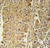 PGD Antibody immunohistochemistry of formalin-fixed and paraffin-embedded human lung carcinoma followed by peroxidase-conjugated secondary antibody and DAB staining.
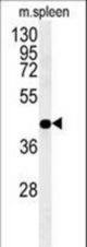 PGD Antibody western blot of mouse spleen tissue lysates (35 ug/lane). The PGD antibody detected PGD protein (arrow).
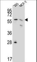 PGD Antibody western blot of 293,MCF-7 cell line lysates (35 ug/lane). The PGD antibody detected PGD protein (arrow).
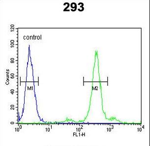 PGD Antibody flow cytometry of 293 cells (right histogram) compared to a negative control cell (left histogram). FITC-conjugated goat-anti-rabbit secondary antibodies were used for the analysis.