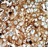
Formalin-fixed and paraffin-embedded human cancer tissue reacted with the primary antibody, which was peroxidase-conjugated to the secondary antibody, followed by AEC staining. This data demonstrates the use of this antibody for immunohistochemistry; clinical relevance has not been evaluated. BC = breast carcinoma; HC = hepatocarcinoma.
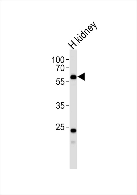
Western blot of lysate from human kidney tissue lysate, using PFKFB3 Antibody (E469). Antibody was diluted at 1:1000. A goat anti-rabbit IgG H&L (HRP) at 1:5000 dilution was used as the secondary antibody. Lysate at 35ug.
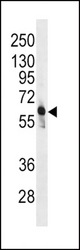
Western blot of anti-PFKFB3 Antibody in CEM cell line lysates (35 ug/lane). PFKFB3(arrow) was detected using the purified antibody.