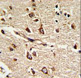
Formalin-fixed and paraffin-embedded human brain tissue reacted with PDIA6 Antibody (Center K159), which was peroxidase-conjugated to the secondary antibody, followed by DAB staining. This data demonstrates the use of this antibody for immunohistochemistry; clinical relevance has not been evaluated.
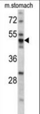
Western blot of PDIA6 antibody (Center K159) in mouse stomach tissue lysates (35 ug/lane). PDIA6 (arrow) was detected using the purified antibody.
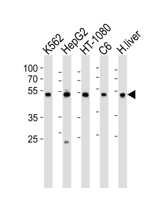
Western blot of lysates from K562, HepG2, HT-1080, rat C6 cell line and human liver tissue lysate (from left to right), using PDIA6 Antibody (Center K159). Antibody was diluted at 1:1000 at each lane. A goat anti-rabbit IgG H&L (HRP) at 1:10000 dilution was used as the secondary antibody. Lysates at 35ug per lane.
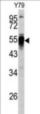
Western blot of PDIA6 antibody (Center K159) in Y79 cell line lysates (35 ug/lane). PDIA6 (arrow) was detected using the purified antibody.
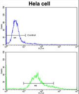
Flow cytometric of HeLa cells using PDIA6 Antibody (Center K159)(bottom histogram) compared to a negative control cell (top histogram)FITC-conjugated goat-anti-rabbit secondary antibodies were used for the analysis.