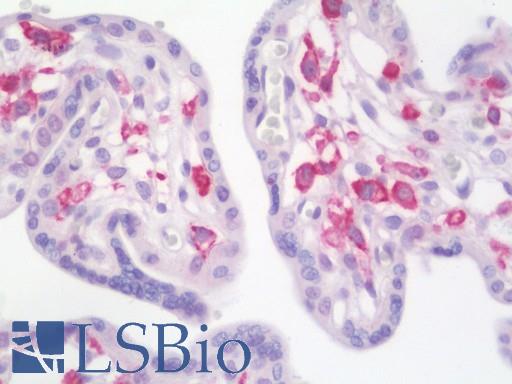
Anti-PDE4B antibody IHC staining of human placenta. Immunohistochemistry of formalin-fixed, paraffin-embedded tissue after heat-induced antigen retrieval. Antibody LS-B11018 concentration 10 ug/ml.
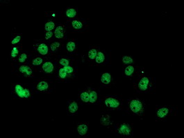
Anti-PDE4B mouse monoclonal antibody immunofluorescent staining of COS7 cells transiently transfected by pCMV6-ENTRY PDE4B.
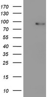
HEK293T cells were transfected with the pCMV6-ENTRY control (Left lane) or pCMV6-ENTRY PDE4B (Right lane) cDNA for 48 hrs and lysed. Equivalent amounts of cell lysates (5 ug per lane) were separated by SDS-PAGE and immunoblotted with anti-PDE4B.

Western blot of extracts (35 ug) from 9 different cell lines by using anti-PDE4B monoclonal antibody (HepG2: human; HeLa: human; SVT2: mouse; A549: human; COS7: monkey; Jurkat: human; MDCK: canine; PC12: rat; MCF7: human).
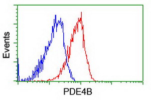
Flow cytometry of HeLa cells, using anti-PDE4B antibody (Red), compared to a nonspecific negative control antibody (Blue).
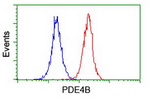
Flow cytometry of Jurkat cells, using anti-PDE4B antibody (Red), compared to a nonspecific negative control antibody (Blue).