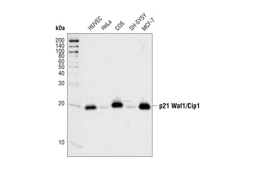
Western blot analysis of extracts from various cell types using p21 Waf1/Cip1 (12D1) Rabbit mAb.

Western blot analysis of extracts from HeLa cells, transfected with 100 nM SignalSilence ® Control siRNA (Fluorescein Conjugate) #6201 (-) or SignalSilence ® p21 Waf1/Cip1 siRNA II (+), using p21 Waf1/Cip1 (12D1) Rabbit mAb #2947 and α-Tubulin (11H10) Rabbit mAb #2125. The p21 Waf1/Cip1 (12D1) Rabbit mAb confirms silencing of p21 Waf1/Cip1 expression and α-Tubulin (11H10) Rabbit mAb is used to control for loading and specificity of p21 Waf1/Cip1 siRNA.
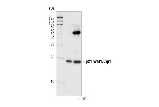
Immunoprecipitation of p21 from human umbillical vein endothelial cells (HUVECs) using p21 Waf1/Cip1 (12D1) Rabbit mAb. Western blot detection was performed using the same antibody.
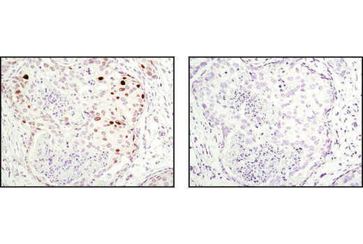
Immunohistochemical analysis of paraffin-embedded human breast carcinoma using p21 Waf1/Cip1 (12D1) Rabbit mAb in the presence of control peptide (left) or antigen-specific peptide (right).
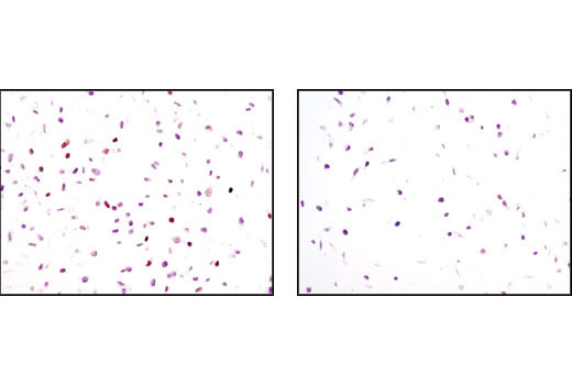
Immunohistochemical analysis of paraffin-embedded HeLa cells, transfected with SignalSilence ® Control siRNA (Unconjugated) #6568 (left) or SignalSilence ® p21 Waf1/Cip1 siRNA II #6558 (right), using p21 Waf1/Cip1 (12D1) Rabbit mAb.
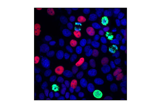
Confocal immunofluorescent analysis of MCF7 cells using p21 Waf1/Cip1 (12D1) Rabbit mAb (red) and Phospho-Histone H3 (Ser10) (6G3) Mouse mAb #9706 (green). Blue pseudocolor = DRAQ5 ® #4084 (fluorescent DNA dye).
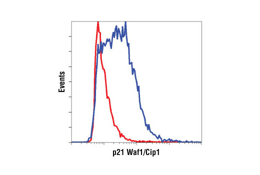
Flow cytometric analysis of HeLa cells (red) and MCF7 cells (blue), using p21 Waf1/Cip1 (12D1) Rabbit mAb.