Anti-OXSM / KS Antibody (clone 1G5)
| Name | Anti-OXSM / KS Antibody (clone 1G5) |
|---|---|
| Supplier | LifeSpan Bioscience |
| Catalog | LS-C175751 |
| Prices | $325.00 |
| Sizes | 100 µl |
| Host | Mouse |
| Clonality | Monoclonal |
| Isotype | IgG2b |
| Clone | 1G5 |
| Applications | IHC-P WB |
| Species Reactivities | Human, Rat |
| Antigen | OXSM / KS antibody was raised against human recombinant protein fragment corresponding to amino acids 78-343 of human OXSM (NP_060367) produced in E. coli. |
| Purity/Format | Protein A/G purified |
| Description | Mouse Monoclonal |
| Gene | OXSM |
| Conjugate | Unconjugated |
| Supplier Page | Shop |
Product images
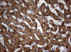 IHC of paraffin-embedded Human liver tissue using anti-OXSM mouse monoclonal antibody.
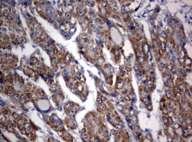 IHC of paraffin-embedded Carcinoma of Human liver tissue using anti-OXSM mouse monoclonal antibody.
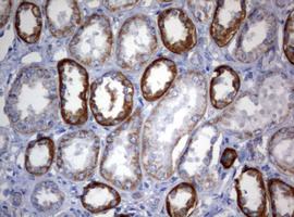 IHC of paraffin-embedded Human Kidney tissue using anti-OXSM mouse monoclonal antibody.
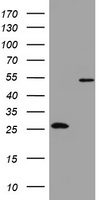 HEK293T cells were transfected with the pCMV6-ENTRY control (Left lane) or pCMV6-ENTRY OXSM (Right lane) cDNA for 48 hrs and lysed. Equivalent amounts of cell lysates (5 ug per lane) were separated by SDS-PAGE and immunoblotted with anti-OXSM.