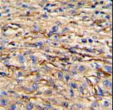
Formalin-fixed and paraffin-embedded human hepatocarcinoma reacted with Nestin Antibody (S1409), which was peroxidase-conjugated to the secondary antibody, followed by DAB staining. This data demonstrates the use of this antibody for immunohistochemistry; clinical relevance has not been evaluated.
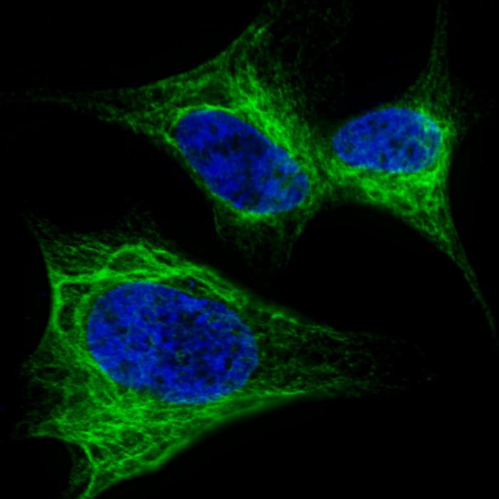
Fluorescent confocal image of SY5Y cells stained with Nestin (S1409) antibody. SY5Y cells were fixed with 4% PFA (20 min), permeabilized with Triton X-100 (0.2%, 30 min). Cells were then incubated Nestin (S1409) primary antibody (1:200, 2 h at room temperature). For secondary antibody, Alexa Fluor 488 conjugated donkey anti-rabbit antibody (green) was used (1:1000, 1h). Nuclei were counterstained with Hoechst 33342 (blue) (10 ug/ml, 5 min). Note the highly specific localization of the Nestin (S1409) immunosignal to the intermediate filaments, supported by Human Protein Atlas Data (http://www.proteinatlas.org/ENSG00000132688).
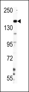
Western blot of Nestin Antibody (S1409) in HepG2 cell line lysates (35 ug/lane). NES (arrow) was detected using the purified antibody.
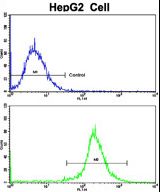
Flow cytometric of HepG2 cells using Nestin Antibody (S1409)(bottom histogram) compared to a negative control cell (top histogram). FITC-conjugated goat-anti-rabbit secondary antibodies were used for the analysis.