
Western blot analysis of extracts from U-87 MG, NCI-H460, A431 cells, and human testis using DAX1 (D2F1) Rabbit mAb. A431 extract is negative as expected.

Immunoprecipitation of DAX1 from KYSE-450 cell extracts using Rabbit (DA1E) mAb IgG XP ® Isotype Control #3900 (lane 2) or DAX1 (D2F1) Rabbit mAb (lane 3). Lane 1 is 10% input. Western blot analysis was performed using DAX1 (D2F1) Rabbit mAb.
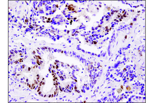
Immunohistochemical analysis of paraffin-embedded human lung carcinoma using DAX1 (D2F1) Rabbit mAb.
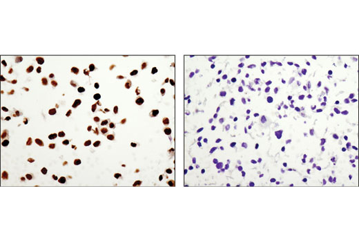
Immunohistochemical analysis of paraffin-embedded NCI-H460 (positive; left) or A431 (negative; right) cell pellets using DAX1 (D2F1) Rabbit mAb.

Immunohistochemical analysis of paraffin-embedded human testis using DAX1 (D2F1) Rabbit mAb.
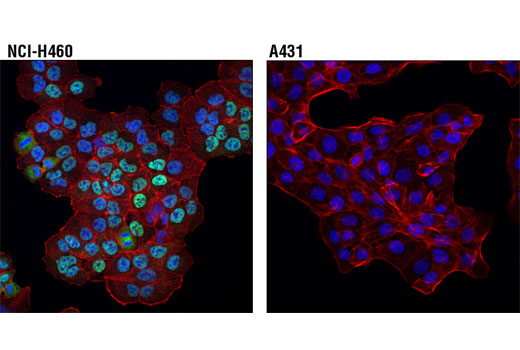
Confocal immunofluorescent analysis of NCI-H460 (positive; upper) and A431 (negative; lower) cells, using DAX1 (D2F1) Rabbit mAb (green). Actin filaments were labeled with DyLight™ 554 Phalloidin #13054 (red). Blue pseudocolor = DRAQ5 ® #4084 (fluorescent DNA dye).