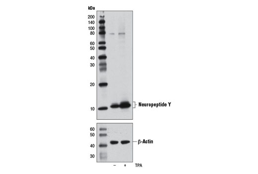
Western blot analysis of extracts from SH-SY5Y cells, untreated (-) or treated with TPA #4174 (200 nM, 24 hr; +), using Neuropeptide Y (D7Y5A) XP ® Rabbit mAb (upper) or β-Actin (D6A8) Rabbit mAb #8457 (lower).

Western blot analysis of the indicated amounts of human peptide YY (PYY), peptide YY (PYY) (3-36), and neuropeptide Y (NPY) peptides using Neuropeptide Y (D7Y5A) XP ® Rabbit mAb (upper). Peptide loading was confirmed by staining overnight with Coomassie Blue (lower).

Western blot analysis of the indicated amounts of human neuropeptide Y using Neuropeptide Y (D7Y5A) XP ® Rabbit mAb.

Western blot analysis of extracts from human hypothalamus and rat spleen using Neuropeptide Y (D7Y5A) XP ® Rabbit mAb.

Immunohistochemical analysis of paraffin-embedded human breast carcinoma using Neuropeptide Y (D7Y5A) XP ® Rabbit mAb.
Immunohistochemical analysis of paraffin-embedded mouse brain using Neuropeptide Y (D7Y5A) XP ® Rabbit mAb.

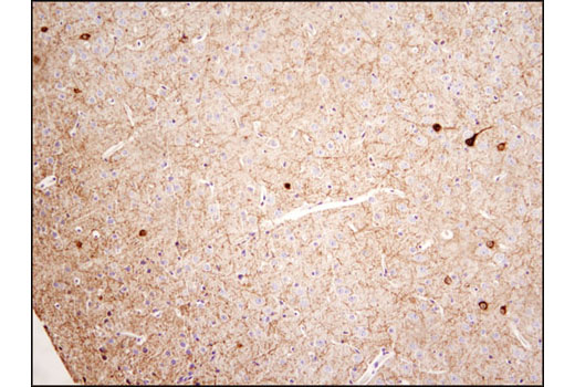
Immunohistochemical analysis of paraffin-embedded rat brain using Neuropeptide Y (D7Y5A) XP ® Rabbit mAb.
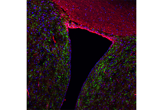
Confocal immunofluorescent analysis of adult mouse hypothalamus using Neuropeptide Y (D7Y5A) XP ® Rabbit mAb (green) and β3-Tubulin (TU-20) Mouse mAb #4466 (red). Blue pseudocolor = DRAQ5 ® #4084 (fluorescent DNA dye).
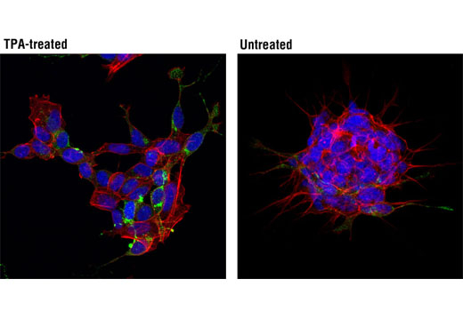
Confocal immunofluorescent analysis of SH-SY5Y cells, treated with TPA #4174 (200 nM, 24 hr; left) or untreated (right), using Neuropeptide Y (D7Y5A) XP ® Rabbit mAb (green). Actin filaments were labeled with DY-554 phalloidin (red). Blue pseudocolor = DRAQ5 ® #4084 (fluorescent DNA dye).