Anti-NRAS / N-ras Antibody (aa72-101)
| Name | Anti-NRAS / N-ras Antibody (aa72-101) |
|---|---|
| Supplier | LifeSpan Bioscience |
| Catalog | LS-C99434 |
| Prices | $295.00 |
| Sizes | 400 µl |
| Host | Rabbit |
| Clonality | Polyclonal |
| Applications | IHC-P WB ELISA |
| Species Reactivities | Human, Mouse, Bat, Bovine, Dog, Horse, Pig |
| Purity/Format | Ammonium sulfate precipitation |
| Blocking Peptide | NRAS / N-ras Antibody Blocking Peptide |
| Description | Rabbit Polyclonal |
| Gene | NRAS |
| Conjugate | Unconjugated |
| Supplier Page | Shop |
Product images
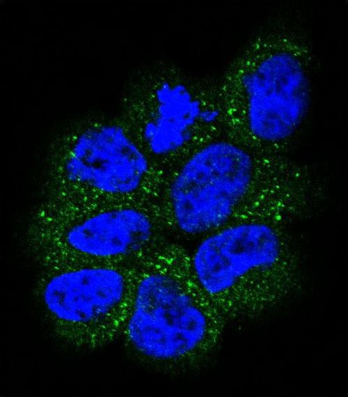 Confocal immunofluorescent of NRAS Antibody with NCI-H460 cell followed by Alexa Fluor 488-conjugated goat anti-rabbit lgG (green). DAPI was used to stain the cell nuclear (blue).
 Western blot of anti-NRAS Antibody in CEM cell line lysates (35 ug/lane). NRAS(arrow) was detected using the purified antibody.
 Western blot of NRAS (arrow) using rabbit polyclonal NRAS Antibody. 293 cell lysates (2 ug/lane) either nontransfected (Lane 1) or transiently transfected with the NRAS gene (Lane 2) (Origene Technologies).
 NRAS Antibody flow cytometry of NCI-H460 cells (right histogram) compared to a negative control cell (left histogram). FITC-conjugated goat-anti-rabbit secondary antibodies were used for the analysis.