Anti-NPPA / ANP Antibody (aa30-56) IHC-plusâ¢
| Name | Anti-NPPA / ANP Antibody (aa30-56) IHC-plus⢠|
|---|---|
| Supplier | LifeSpan Bioscience |
| Catalog | LS-B9895 |
| Prices | $395.00 |
| Sizes | 200 µl |
| Host | Rabbit |
| Clonality | Polyclonal |
| Applications | IHC-P RIA |
| Species Reactivities | Human |
| Purity/Format | Protein A purified |
| Blocking Peptide | NPHS1 / Nephrin Antibody Blocking Peptide |
| Description | Rabbit Polyclonal |
| Gene | NPPA |
| Conjugate | Unconjugated |
| Supplier Page | Shop |
Product images
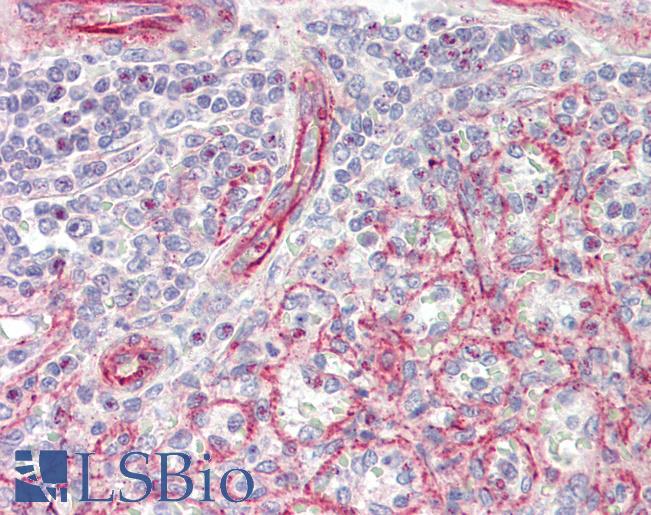 Anti-NPPA / ANP antibody IHC staining of human spleen. Immunohistochemistry of formalin-fixed, paraffin-embedded tissue after heat-induced antigen retrieval.
 Western blot of lysate from mouse heart tissue lysate, using NPPA Antibody. Antibody was diluted at 1:1000. A goat anti-rabbit IgG H&L (HRP) at 1:5000 dilution was used as the secondary antibody. Lysate at 35ug.
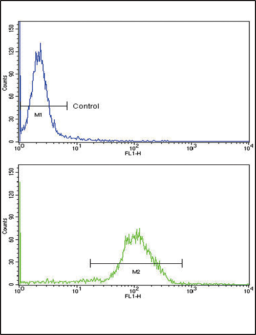 Flow cytometric of MDA-231 cells using NPPA Antibody (bottom histogram) compared to a negative control cell (top histogram). FITC-conjugated goat-anti-rabbit secondary antibodies were used for the analysis.