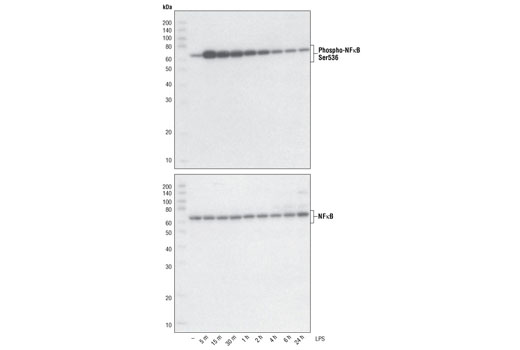
Western blot analysis of extracts from THP-1 cells, differentiated with TPA (#9905, 80 nM for 24h) and treated with 1 μg/ml LPS for the indicated times, using Phospho-NF-κB p65 (Ser536) (93H1) Rabbit mAb #3033 (upper) and NF-κB p65 (C22B4) Rabbit mAb (lower).

Western blot analysis of extracts from HeLa (human), PC12 (rat) and Neuro2A (mouse) cell lines using NF-κB p65 (C22B4) Rabbit mAb.

Western blot analysis of extracts from HeLa cells, transfected with 100 nM SignalSilence ® Control siRNA (Fluorescein Conjugate) #6201 (-), SignalSilence ® NF-κB p65 siRNA II (+), or SignalSilence® NF-κB p65 siRNA I #6261 (+), using NF-κB p65 (C22B4) Rabbit mAb #4764 and α-Tubulin (11H10) Rabbit mAb #2125. NF-κB p65 (C22B4) Rabbit mAb confirms silencing of NF-κB p65 and α-Tubulin (11H10) Rabbit mAb is used to control for loading and specificity of NF-κB p65 siRNA.

Confocal immunofluorescent analysis of HeLa cells, untreated (left) or TNF-α-treated (#8902, 20ng/ml for 20 min, right), using NF-κB p65 (C22B4) Rabbit mAb (green). Actin filaments have been labeled with Alexa Fluor ® 555 phalloidin (red). Blue pseudocolor = DRAQ5™ (fluorescent DNA dye).

Flow cytometric analysis of OVCAR8 cells using NF-κB p65 (C22B4) Rabbit mAb (blue) compared to a nonspecific negative control antibody (red).