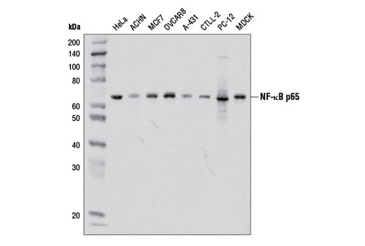
Western blot analysis of extracts from various cell lines using NF-κB p65 (D14E12) XP ® Rabbit mAb.

Immunohistochemical analysis using NF-κB p65 (D14E12) XP ® Rabbit mAb on SignalSlide ® NF-κB p65 IHC Controls #12873 (paraffin-embedded HCT116 cells, untreated (left) or treated with hTNF-α #8902 (right)).

Immunohistochemical analysis of paraffin-embedded human chronic cholecystitis using NF-κB p65 (D14E12) XP ® Rabbit mAb.

Confocal immunofluorescent analysis of HT-1080 cells, untreated (left) or treated with hTNF-α #8902 (20 ng/ml, 20 min) (right), using NF-κB p65 (D14E12) XP ® Rabbit mAb (green). Actin filaments were labeled with DY-554 phalloidin (red). Blue pseudocolor = DRAQ5 ® #4084 (fluorescent DNA dye).

Flow cytometric analysis of HeLa cells using NF-κB p65 (D14E12) XP ® Rabbit mAb (blue) compared to concentration matched Rabbit (DA1E) mAb IgG XP ® Isotype Control #3900 (red).

Chromatin immunoprecipitations were performed with cross-linked chromatin from 4 x 10 6 HeLa cells treated with hTNF-α #8902 (30 ng/ml, 1 hr) and 5 μl of NF-κB p65 (D14E12) XP ® Rabbit mAb, using SimpleChIP ® Enzymatic Chromatin IP Kit (Magnetic Beads) #9005. DNA Libraries were prepared from 5 ng enriched ChIP DNA using NEBNext ® Ultra™ II DNA Library Prep Kit for Illumina ® , and sequenced on the Illumina NextSeq. The figure shows binding across IL-8, a known target gene of NFκB (see additional figure containing ChIP-qPCR data). For additional ChIP-seq tracks, please download the product data sheet.

Chromatin immunoprecipitations were performed with cross-linked chromatin from 4 x 10 6 HeLa cells treated with hTNF-α #8902 (30 ng/ml, 1 hr) and either 5 μl of NF-κB p65 (D14E12) XP ® Rabbit mAb or 2 μl of Normal Rabbit IgG #2729 using SimpleChIP ® Enzymatic Chromatin IP Kit (Magnetic Beads) #9003. The enriched DNA was quantified by Real-Time PCR using SimpleChIP ® Human IκBα Promoter Primers #5552, human IL-8 promoter primers, and SimpleChIP ® Human α Satellite Repeat Primers #4486. The amount of immunoprecipitated DNA in each sample is represented as signal relative to the total amount of input chromatin, which is equivalent to one.