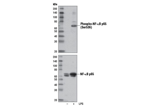
Western blot analysis of extracts from THP-1 cells differentiated with TPA #4174 (80 nM, overnight), untreated (-) or LPS-treated (1 μg/ml, 15 min; +), using Phospho-NF-κB p65 (Ser536) (E1Z1T) Mouse mAb (upper) or NF-κB p65 (D14E12) XP ® Rabbit mAb #8242 (lower).

Western blot analysis of extracts from HeLa and PC-3 cells, untreated (-) or treated with Human Tumor Necrosis Factor-α (hTNF-α) #8902 (hTNF-α; 20 ng/ml, 5 min; +), using Phospho-NF-κB p65 (Ser536) (E1Z1T) Mouse mAb (upper) or NF-κB p65 (D14E12) XP ® Rabbit mAb #8242 (lower).

Immunoprecipitation of phospho-NF-κB p65 (Ser536) from HeLa cells treated with Human Tumor Necrosis Factor-α (hTNF-α) #8902 (20 ng/ml, 5 min) using Mouse (G3A1) mAb IgG1 Isotype Control #5415 (lane 2) or Phospho-NF-κB p65 (Ser536) (E1Z1T) Mouse mAb (lane 3). Lane 1 represents 10% input. Western blot was performed using Phospho-NF-κB p65 (Ser536) (93H1) Rabbit mAb #3033.