MUC1 (D9O8K) XP ® Rabbit mAb
| Name | MUC1 (D9O8K) XP ® Rabbit mAb |
|---|---|
| Supplier | Cell Signaling Technology |
| Catalog | 14161 |
| Prices | $109.00, $277.00 |
| Sizes | 20 µl (2 western blots), 100 µl (10 western blots) |
| Host | Rabbit |
| Clonality | Monoclonal |
| Isotype | IgG |
| Clone | D9O8K |
| Applications | WB IP ICC/IF FC |
| Species Reactivities | Human |
| Antigen | Monoclonal antibody is produced by immunizing animals with a synthetic peptide corresponding to residues near the amino terminus of human MUC1 protein. |
| Description | Rabbit Monoclonal |
| Gene | MUC1 |
| Supplier Page | Shop |
Product images
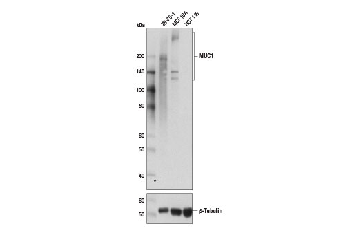 Western blot analysis of extracts from ZR-75-1, MCF 10A, and HCT 116 cells using MUC1 (D9O8K) XP ® Rabbit mAb (upper) and β-Tubulin Antibody #2146 (lower).
 Confocal immunofluorescent analysis of ZR-75-1 (positive, left) and HCT 116 (negative, right) cells using MUC1 (D9O8K) XP ® Rabbit mAb (green). Blue pseudocolor = DRAQ5 ® #4084 (fluorescent DNA dye).
 Flow cytometric analysis of HCT 116 (blue) and ZR-75-1 (green) using MUC1 (D9O8K) XP ® Rabbit mAb. Anti-rabbit IgG (H+L), F(ab') 2 Fragment (Alexa Fluor ® 488 Conjugate) #4412 was used as a secondary antibody.