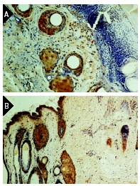
Immunoperoxidase staining of formalin-fixed, paraffin-embedded wounded ovine skin one day following excisional injury. Sections probed with TGFβ RII (C-16): sc-220 (A) and TGFβ RII (L-21): sc-400 (B). Kindly provided by Leslie Gold.
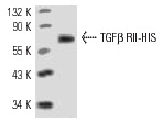
TGFβ RII (C-16): sc-220. Western blot analysis of HIS-tagged human recombinant TGFβ RII.
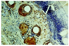
Immunoperoxidase staining of formalin-fixed, paraffin-embedded wounded ovine skin one day following excisional injury. Sections probed with TGFβ RII (C-16): sc-220 (A) and TGFβ RII (L-21): sc-400 (B). Kindly provided by Leslie Gold.
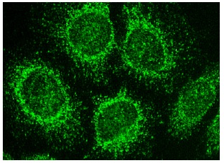
TGFβ RII (C-16): sc-220. Immunofluorescence staining of methanol-fixed HeLa cells showing cytoplasmic localization.