
Western blot analysis of PDGFR-β expression in rodent 3611-RF (A,C) and human CCD-1064Sk (B,D) fibroblast cell lines. Antibodies tested include PDGFR-β (P-20)-G: sc-339-G (A,B) and PDGFR-β (M-20): sc-1627 (C,D).
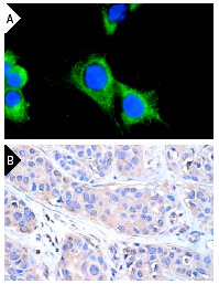
PDGFR-β (P-20): sc-339. Immunofluorescence staining of methanol-fixed NIH/3T3 cells (A). Note membrane and cytoplasmic fluorescein immunostaining and nuclear DAPI counterstain. Immunoperoxidase staining of formalin-fixed, paraffin-embedded human breast carcinoma tissue at high magnification (B) showing membrane and cytoplasmic localization.
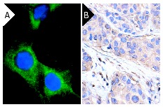
PDGFR-β (P-20): sc-339. Immunofluorescence staining of methanol-fixed NIH/3T3 cells (A). Note membrane and cytoplasmic fluorescein immunostaining and nuclear DAPI counterstain. Immunoperoxidase staining of formalin-fixed, paraffin-embedded human breast carcinoma tissue at high magnification (B) showing membrane and cytoplasmic localization.
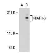
PDGFR-β (P-20): sc-339. Western blot analysis of PDGFR-β expression in non-transfected: sc-117752 (A) and human PDGFR-β transfected: sc-114235 (B) 293T whole cell lysates.
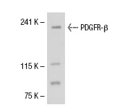
PDGFR-β (P-20): sc-339. Western blot analysis of PDGFR-β expression in CCD-1064Sk whole cell lysate.
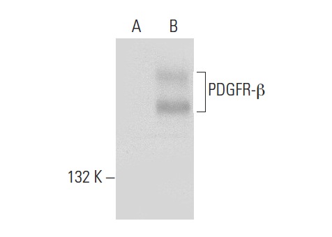
PDGFR-β (P-20)-G: sc-339-G. Western blot analysis of PDGFR-β expression in non-transfected: sc-117752 (A) and human PDGFR-β transfected: sc-159386 (B) 293T whole cell lysates.