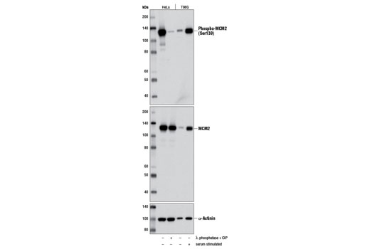
Western blot analysis of HeLa cell extracts, untreated (-) or calf intestinal phosphatase (CIP) and λ phosphatase-treated (+), and extracts of T98G cells serum-starved for 48 hours (-) or serum starved for 48 hours followed by serum-stimulation for 24 hours (+), using Phospho-MCM2 (Ser139) (D1Z8X) XP ® Rabbit mAb (upper), MCM2 (D7G11) XP ® Rabbit mAb #3619 or α-Actinin (D6F6) XP ® Rabbit mAb #6487.
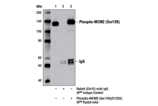
Immunoprecipitation of phospho-MCM2 (Ser139) from T98G cell extracts, using Rabbit (DA1E) mAb IgG XP ® Isotype Control #3900 (lane 2) or Phospho-MCM2 (Ser139) (D1Z8X) XP ® Rabbit mAb (lane 3). Lane 1 is 10% input. Western blot analysis was performed using Phospho-MCM2 (Ser139) (D1Z8X) XP ® Rabbit mAb.
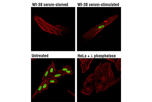
Confocal immunofluorescent analysis of WI-38 cells, serum-deprived (48 hr; upper left), or serum-treated (10%, 24 hr; upper right) and HeLa cells, untreated (lower left), or λ phosphatase-treated (2 hr; lower right), using Phospho-MCM2 (Ser139) (D1Z8X) XP ® Rabbit mAb (green). Actin filaments were labeled with DY-554 phalloidin (red).
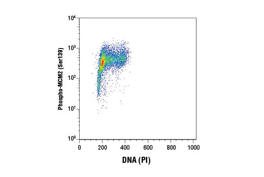
Flow cytometric analysis of Jurkat cells using Phospho-MCM2 (Ser139) (D1Z8X) XP ® Rabbit mAb and Propidium Iodide (PI)/RNase Staining Solution #4087 (DNA content). Anti-rabbit IgG (H+L), F(ab') 2 Fragment (Alexa Fluor ® 488 conjugate) #4412 was used as a secondary Ab.