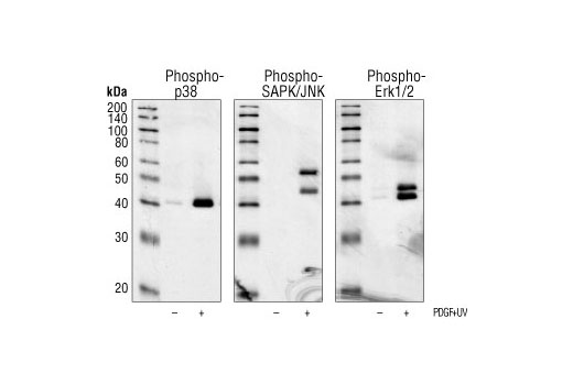
Specificity of Phospho-Erk1/2, Phospho-p38 MAPK and Phospho-SAPK/JNK Rabbit mAb: Western blot analysis of extracts from NIH/3T3 cells treated with PDGF and UV, using Phospho-p38 MAPK Rabbit mAb #9215, Phospho-SAPK/JNK Rabbit mAb and Phospho-Erk1/2 Rabbit mAb.

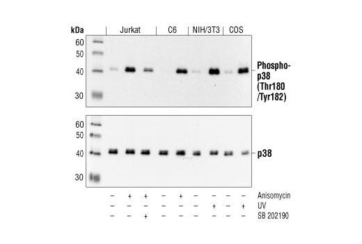
Western blot analysis of extracts from Jurkat, C6, NIH/3T3 and COS cells, untreated or treated as indicated, using Phospho-p38 MAPK (Thr180/Tyr182) (3D7) Rabbit mAb (upper) or p38 MAPK Antibody #9212 (lower).

Confocal immunofluorescent analysis of HeLa cells, untreated (left) or anisomycin-treated (right), using Phospho-p38 MAPK (Thr180/Tyr182)(3D7) Rabbit mAb (green). Actin filaments have been labeled with Alexa Fluor® 555 phalloidin (red).

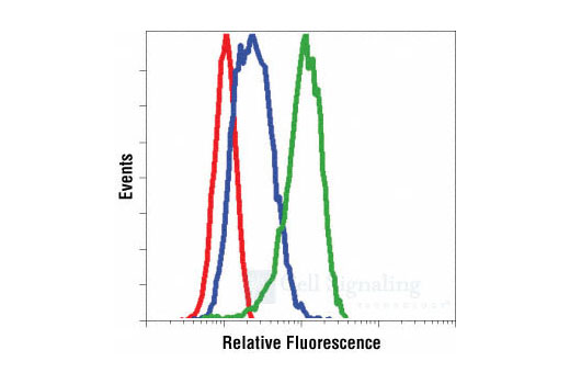
Flow cytometric analysis of Jurkat cells, untreated (blue) or anisomycin-treated (green), using Phospho-p38 MAPK (Thr180/Tyr182) (3D7) Rabbit mAb compared to a nonspecific negative control antibody (red).


