Endoglin (P3D1)
| Name | Endoglin (P3D1) |
|---|---|
| Supplier | Santa Cruz Biotechnology |
| Catalog | sc-18838 |
| Prices | $279.00 |
| Sizes | 200 µg/ml |
| Host | Mouse |
| Clonality | Monoclonal |
| Isotype | mouse IgG 1 |
| Applications | WB IP ICC/IF FC |
| Species Reactivities | Mouse, Human |
| Antigen | FL (h) |
| Description | Mouse Monoclonal |
| Gene | ENG |
| Supplier Page | Shop |
Product images
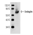 Endoglin (P3D1): sc-18838. Western blot analysis of Endoglin expression in mouse embryo tissue extract.
 Endoglin (P3D1) PE: sc-18838 PE. FCM analysis of PMA-stimulated TF-1 + PMA cells. Black line histogram represents the isotype control, normal mouse IgG.
 Endoglin (P3D1) FITC: sc-18838 FITC. FCM analysis of PMA-stimulated TF-1 + PMA cells. Black line histogram represents the isotype control, normal mouse IgG.
 Endoglin (P3D1): sc-18838. Immunofluorescence staining of methanol-fixed HUV-EC-C cells showing membrane localization.