Anti-IP6K1 Antibody (aa50-309)
| Name | Anti-IP6K1 Antibody (aa50-309) |
|---|---|
| Supplier | LifeSpan Bioscience |
| Catalog | LS-C185578 |
| Prices | $335.00 |
| Sizes | 100 µl |
| Host | Rabbit |
| Clonality | Polyclonal |
| Isotype | IgG |
| Applications | IHC-P ICC/IF WB |
| Species Reactivities | Human |
| Antigen | IP6K1 antibody was raised against recombinant fragment corresponding to a region within amino acids 50 and 309 of IP6K1 (SwissProt Q92551). |
| Purity/Format | Immunoaffinity purified |
| Description | Rabbit Polyclonal |
| Gene | IP6K1 |
| Conjugate | Unconjugated |
| Supplier Page | Shop |
Product images
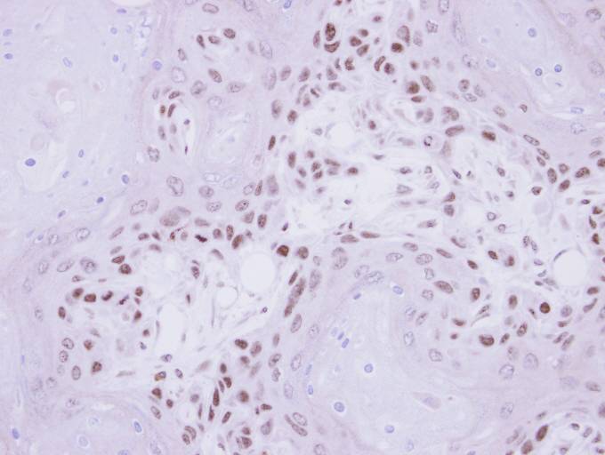 IHC of paraffin-embedded Ca922 Xenograft using IP6K1 antibody at 1:500 dilution.
 Confocal immunofluorescence analysis (Olympus FV10i) of paraformaldehyde-fixed HeLa using IP6K1 antibody (Green) at 1:500 dilution. Alpha-tubulin filaments were labeled with alpha-tubulin antibody (Red) at 1:2000.
 IP6K1 antibody detects IHPK1 protein by Western blot analysis. A. 30 ug 293T whole cell lysate/extract. B. 30 ug whole cell lysate/extract of human IHPK1-transfected 293T cells. 10 % SDS-PAGE. IP6K1 antibody dilution:1:5000