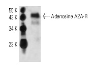
Adenosine A2A-R (7F6-G5-A2): sc-32261. Western blot analysis of Adenosine A2A-R expression in mouse brain tissue extract.

Adenosine A2A-R (7F6-G5-A2) PE: sc-32261 PE. FCM analysis of human peripheral blood leukocytes. Black line histogram represents the isotype control, normal mouse IgG
2a: sc-2867.
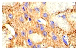
Adenosine A2A-R (7F6-G5-A2): sc-32261. Immunoperoxidase staining of formalin fixed, paraffin-embedded mouse brain tissue showing membrane localization.
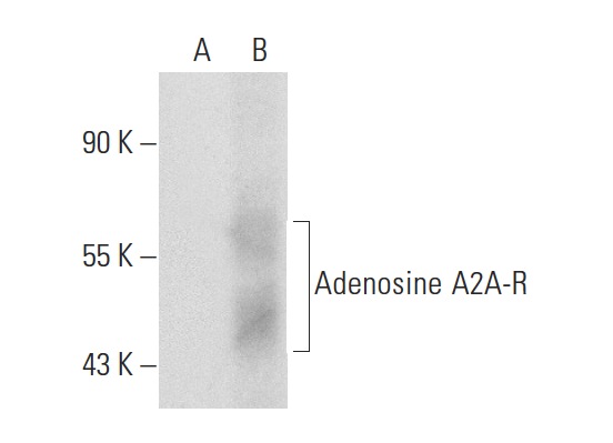
Adenosine A2A-R (7F6-G5-A2): sc-32261. Western blot analysis of Adenosine A2A-R expression in non-transfected: sc-117752 (A) and human Adenosine A2A-R transfected: sc-127942 (B) 293T whole cell lysates.