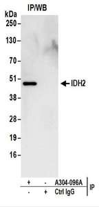
Detection of Human IDH2 by Western Blot of Immunoprecipitates. Samples: Whole cell lysate (1 mg for IP; 20% of IP loaded) from 293T cells. Antibodies: Affinity purified rabbit anti-IDH2 antibody used for IP at 6 ug/mg lysate. For blotting immunoprecipitated IDH2, was used at 1 ug/ml. Detection: Chemiluminescence with an exposure time of 10 seconds.
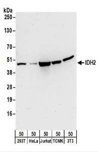
Detection of Human and Mouse IDH2 by Western Blot. Samples: Whole cell lysate (50 ug) from 293T, HeLa, Jurkat, mouse TCMK-1, and mouse NIH3T3 cells. Antibodies: Affinity purified rabbit anti-IDH2 antibody used for WB at 0.4 ug/ml. Detection: Chemiluminescence with an exposure time of 30 seconds.