TGFβ1 (V)
| Name | TGFβ1 (V) |
|---|---|
| Supplier | Santa Cruz Biotechnology |
| Catalog | sc-146 |
| Prices | $279.00 |
| Sizes | 200 µg/ml |
| Host | Rabbit |
| Clonality | Polyclonal |
| Isotype | rabbit IgG |
| Applications | WB IP ICC/IF IHC-P ELISA |
| Species Reactivities | Mouse, Rat, Human, Horse, Bovine, Pig, Dog, Arabidopsis thaliana, Xenopus |
| Antigen | C-terminal (h) |
| Description | Rabbit Polyclonal |
| Gene | TGFB1 |
| Supplier Page | Shop |
Product images
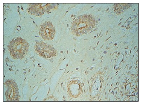 TGFβ1 (V): sc-146. Immunoperoxidase staining of formalin fixed, paraffin-embedded porcine uterus tissue. Kindly provided by Laurie A. Jaeger.
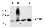 TGFβ1 (V)-G: sc-146-G. Western blot analysis of TGFβ1 expression in rat adrenal (A) and mouse uterus (B) extracts and MCF7 (C) and T-47D (D) whole cell lysates.
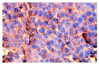 TGFβ1 (V): sc-146. Immunoperoxidase staining of formalin fixed, paraffin-embedded mouse ovary tissue showing extracellular localization.
 TGFβ1 (V): sc-146. Western blot analysis of TGFβ1 expression in 293T whole cell lysate.
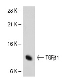
 TGFβ1 (V): sc-146. Western blot analysis of TGFβ1 expression in rat adrenal gland (A) and mouse uterus (B) tissue extracts.