Anti-HSD2 / HSD11B2 Antibody (aa277-306) IHC-plusâ¢
| Name | Anti-HSD2 / HSD11B2 Antibody (aa277-306) IHC-plus⢠|
|---|---|
| Supplier | LifeSpan Bioscience |
| Catalog | LS-B10555 |
| Prices | $395.00 |
| Sizes | 200 µl |
| Host | Rabbit |
| Clonality | Polyclonal |
| Applications | WB ELISA |
| Species Reactivities | Rat |
| Purity/Format | Protein A purified |
| Blocking Peptide | HSD17B12 Antibody Blocking Peptide |
| Description | Rabbit Polyclonal |
| Gene | HSD11B2 |
| Conjugate | Unconjugated |
| Supplier Page | Shop |
Product images
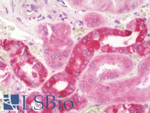 Anti-HSD2 / HSD11B2 antibody IHC staining of human kidney. Immunohistochemistry of formalin-fixed, paraffin-embedded tissue after heat-induced antigen retrieval. Antibody LS-B10555 dilution 1:100.
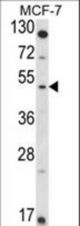 Western blot of HSD11B2 Antibody in MCF-7 cell line lysates (35 ug/lane). HSD11B2 (arrow) was detected using the purified antibody.
 HSD11B2 Antibody flow cytometry of MCF-7 cells (bottom histogram) compared to a negative control cell (top histogram). FITC-conjugated goat-anti-rabbit secondary antibodies were used for the analysis.