ITI-H2 (K-16)
| Name | ITI-H2 (K-16) |
|---|---|
| Supplier | Santa Cruz Biotechnology |
| Catalog | sc-21975 |
| Prices | $279.00 |
| Sizes | 200 µg/ml |
| Host | Goat |
| Clonality | Polyclonal |
| Isotype | goat IgG |
| Applications | WB IP ICC/IF IHC-P ELISA |
| Species Reactivities | Human |
| Antigen | internal (h) |
| Description | Goat Polyclonal |
| Gene | ITIH2 |
| Supplier Page | Shop |
Product images
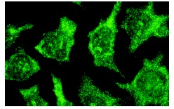 ITI-H2 (K-16): sc-21975. Immunofluorescence staining of methanol-fixed HeLa cells showing cell surface localization.
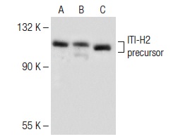 ITI-H2 (K-16): sc-21975. Western blot analysis of ITI-H2 expression in RAW 264.7 (A), HeLa (B) and CTLL-2 (C) whole cell lysates.
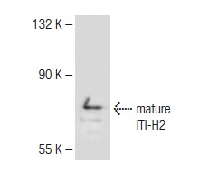 ITI-H2 (K-16): sc-21975. Western blot analysis of ITI-H2 expression in Jurkat whole cell lysate.
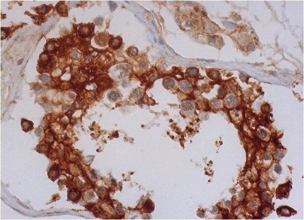 ITI-H2 (K-16): sc-21975. Immunoperoxidase staining of formalin fixed, paraffin-embedded human testis tissue showing membrane and cytoplasmic staining of glandular cells and Leydig cells.