Anti-HLA-DQA1 Antibody (aa39-66)
| Name | Anti-HLA-DQA1 Antibody (aa39-66) |
|---|---|
| Supplier | LifeSpan Bioscience |
| Catalog | LS-C164916 |
| Prices | $295.00 |
| Sizes | 400 µl |
| Host | Rabbit |
| Clonality | Polyclonal |
| Applications | WB ELISA |
| Species Reactivities | Human |
| Purity/Format | Protein A purified |
| Blocking Peptide | HLA-C Antibody Blocking Peptide |
| Description | Rabbit Polyclonal |
| Gene | HLA-DQA1 |
| Conjugate | Unconjugated |
| Supplier Page | Shop |
Product images
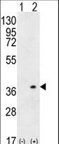 Western blot of HLA-DQA1 (arrow) using rabbit polyclonal HLA-DQA1 Antibody. 293 cell lysates (2 ug/lane) either nontransfected (Lane 1) or transiently transfected (Lane 2) with the HLA-DQA1 gene.
 HLA-DQA1 Antibody western blot of NCI-H460 cell line lysates (35 ug/lane). The HLA-DQA1 antibody detected the HLA-DQA1 protein (arrow).
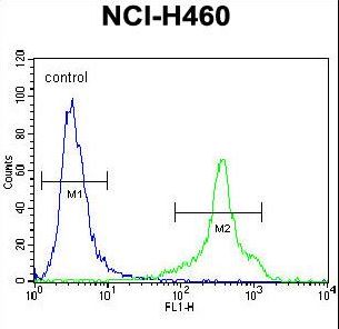 HLA-DQA1 Antibody flow cytometry of NCI-H460 cells (right histogram) compared to a negative control cell (left histogram). FITC-conjugated goat-anti-rabbit secondary antibodies were used for the analysis.